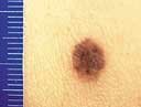
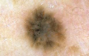
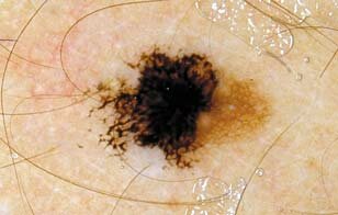
This melanocytic proliferation belongs to the “gray zone” because dermoscopic as well as histopathologic criteria for the diagnosis of Clark nevus and melanoma in situ are both present.
|
|
|
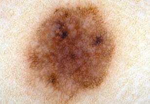
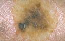
Dermoscopically the diagnosis of this melanocytic proliferation is suggestive of melanoma, albeit different histopathologists came to different diagnostic conclusions. Therefore we interpreted this lesion as belonging to the “gray zone”.
|
|
|
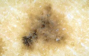
This melanocytic proliferation reveals dermoscopic and histopathologic criteria for both Clark nevus and melanoma in situ. Thus we categorized this lesion within the “gray zone”.
|

|
|
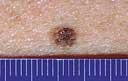
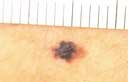
Dermoscopically this melanocytic proliferation with peripheral hyperpigmentation due to an atypical pigment network is suggestive of melanoma in situ. However, the histopathologic diagnosis performed by different histopathologists was equivocal.
|
|
|
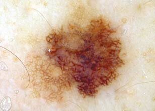
This melanocytic proliferation has many features of melanoma in situ. However, the diagnosis of melanoma in situ has not been confirmed by all the histopathologists studying this case.
|
|
|
)
)
)
)
)
)
)
)
)
)